

Quick answer
A kidney cyst is a common finding, and a simple cyst is benign. The Bosniak classification sorts cysts by appearance and guides what to do: classes I and II need no follow-up, IIF is followed with imaging, III calls for surgery or active surveillance, and IV calls for surgery. A solid mass is a different situation, and about 15% of kidney tumors are benign.
Three different findings
The report of an ultrasound or CT done for another reason often mentions a lesion in the kidney. The words in the report make a difference.
Simple cyst
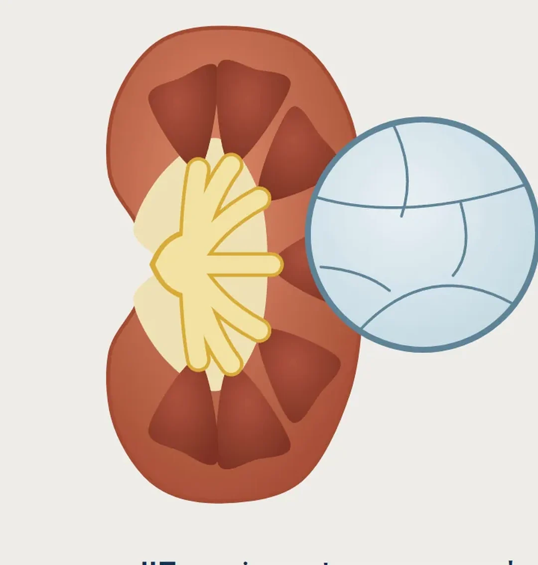
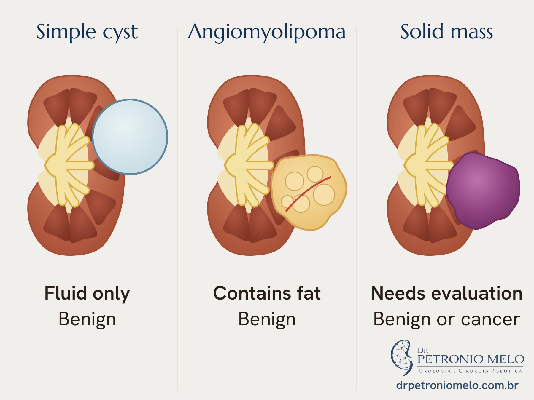
A simple cyst is a fluid-filled sac with a thin wall. In a study of people who had routine check-ups, it appeared in 7.7%: 2.7% under age 40 and 23.9% over 60.5
Bosniak classification
The classification uses the appearance of the cyst on CT or MRI with contrast. The 2019 version defined each class more precisely.3
| Class | Appearance | Approach in the European guideline |
|---|---|---|
| I | Thin wall, fluid only | Benign, no follow-up2 |
| II | 1 to 3 thin septa | Benign, no follow-up2 |
| IIF | More septa, or a slightly thicker wall | Follow-up for up to five years2 |
| III | Thick or irregular wall or septa | Surgery or active surveillance2 |
| IV | Nodule that enhances with contrast | Surgery2 |
In the groups gathered by the guideline, the proportion of cancer was 51% in class III and 89% in class IV.1 In a 2025 review, the proportion in class IIF changed with the way the result was confirmed: 2% when the reference was imaging follow-up and 41% when it was pathology.6
For complex cysts, MRI may be preferable to CT.2 Biopsy is not recommended for cysts, unless there are solid areas.2
Angiomyolipoma
It is a benign tumor, found in about 0.44% of people, more often in women.1 The fat inside it usually allows the diagnosis on imaging. When there is little fat, imaging may not be conclusive.1
Monitoring is the most common approach. In a group of patients followed over time, 11% of tumors grew, 2% bled and 5% needed treatment.1 The European guideline says the traditional 4 cm threshold, on its own, should not lead to treatment.1
Treatment is recommended if there is persistent pain, bleeding or a very large tumor.1 When surgery is needed, the preference is to preserve the kidney. Embolization, which blocks the tumor's blood vessels, is less invasive but needs repeat treatment more often: 31% against 0.85% with surgery.1,4
Solid mass
A solid mass calls for evaluation by a urologist. About 15% of kidney tumors are benign.1 Oncocytoma is one example: it is benign and accounts for 4% to 7% of solid tumors, but imaging cannot reliably tell it apart from cancer, and needle biopsy does not settle this diagnosis.1,2
Biopsy can help in some cases. In a study of 542 small masses that were operated on, a benign finding at surgery was less frequent in centers that did biopsies: 5% against 16%.2
When the evaluation suggests cancer, the options are on the kidney cancer page. In many cases, only the tumor can be removed, with robotic partial nephrectomy.
How I approach it
In my practice, I read the images together with the report and explain which of these groups the lesion belongs to. A benign cyst does not need to become a worry. When there is doubt, we assess together whether a better test, follow-up or treatment is worthwhile, and your family takes part if you wish.
When to see a urologist
- If the report mentions a complex cyst, Bosniak class IIF, III or IV, or a solid mass
- If the report mentions an angiomyolipoma
- If blood appears in your urine
- If you have flank pain that does not go away
Frequently asked questions
Is a kidney cyst dangerous?+
A simple cyst is benign. Bosniak classes I and II do not need follow-up.1 Cysts with a thick wall, thick septa or a nodule carry a higher risk of cancer and need evaluation.1,2
Can a kidney cyst turn into cancer?+
In a review cited by the European guideline, under 1% of stable IIF cysts showed cancer during follow-up, and 12% changed to III or IV.1 That is why class IIF is followed with imaging for up to five years.2
What is an angiomyolipoma?+
It is a benign kidney tumor made of fat, muscle and blood vessels.1,4 It is often just monitored. Treatment is recommended if there is persistent pain, bleeding or a very large tumor.1
Is every solid kidney mass cancer?+
No. About 15% of kidney tumors are benign.1 Imaging cannot always tell a benign mass from a cancer, and the plan depends on each case.2
Does a kidney cyst need a biopsy?+
Usually not. The European guideline does not recommend biopsy of cysts, unless there are solid areas, as in class IV.2
References
- EAU Guidelines on Renal Cell Carcinoma. European Association of Urology; 2026. Epidemiology, aetiology and pathology chapter. EAU
- EAU Guidelines on Renal Cell Carcinoma. European Association of Urology; 2026. Diagnostic evaluation chapter. EAU
- Silverman SG, Pedrosa I, Ellis JH, et al. Bosniak Classification of Cystic Renal Masses, Version 2019: An Update Proposal and Needs Assessment. Radiology. 2019;292(2):475-488. PubMed
- Fernández-Pello S, Hora M, Kuusk T, et al. Management of Sporadic Renal Angiomyolipomas: A Systematic Review of Available Evidence to Guide Recommendations from the European Association of Urology Renal Cell Carcinoma Guidelines Panel. Eur Urol Oncol. 2020;3(1):57-72. PubMed
- Ozveren B, Onganer E, Türkeri LN. Simple Renal Cysts: Prevalence, Associated Risk Factors and Follow-Up in a Health Screening Cohort. Urol J. 2016;13(1):2569-75. PubMed
- McGrath TA, Davenport MS, Silverman SG, et al. Bosniak Classification of Cystic Renal Masses Version 2019: Proportion of Malignancy by Class and Subclass-Systematic Review and Meta-Analysis. AJR Am J Roentgenol. 2025;224(3):e2432342. PubMed
Would you like to talk about your case?
In the consultation, I go over your tests and your priorities.
Book a consultation