

Quick answer
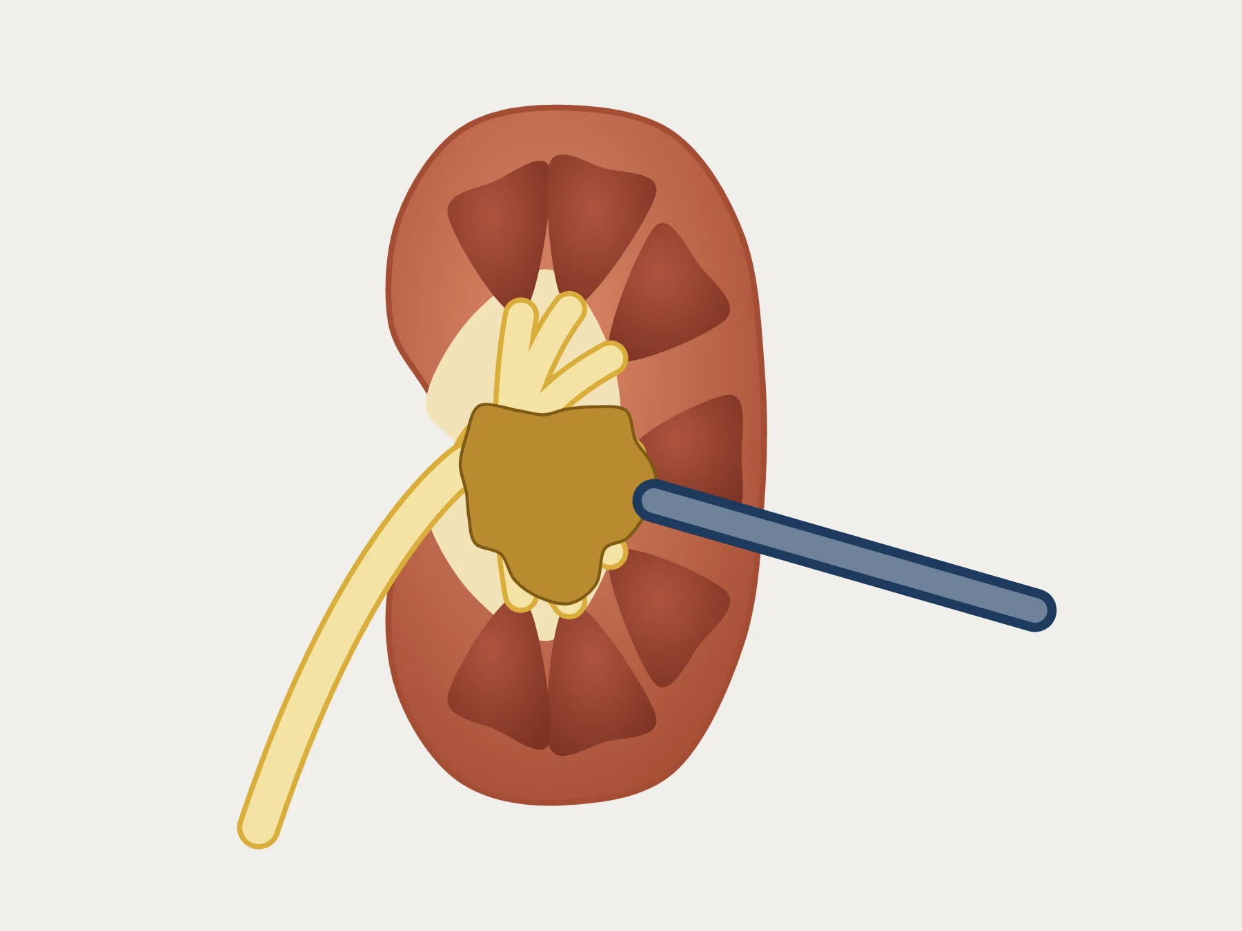
Percutaneous nephrolithotomy is surgery in which a small access, through the side or the back, reaches the kidney to break up and remove the stone. It is the first option for kidney stones above 2 cm.1 In my practice, I always operate with the patient face up, without a nephrostomy tube whenever possible, and the hospital stay is 24 hours.
How it is done
- PlanningThe CT scan shows the stone, the inside of the kidney and the organs around it, which makes the access safer.1
- PositionIn my practice, the patient is always face up, usually in the Barts flank-free position.
- PunctureA needle goes through the skin and reaches a calyx of the kidney, guided by fluoroscopy, ultrasound or both.1
- DilationThe track is widened and a sheath stays in place, forming a tunnel to the kidney.1
- Breaking and removalThe nephroscope goes in through the tunnel. The stone is broken up with ultrasonic, pneumatic or laser energy, and the fragments are removed.1
- EndWhenever possible, I finish without a nephrostomy tube, with only the double J stent. The hospital stay is 24 hours.
Patient position
The operation can be done face down or face up. The European guideline considers both equally safe, with the same results.1 In a meta-analysis of 11,774 patients, the face-up position had a shorter operating time and fewer serious complications.1 It also allows a flexible ureteroscope to be used through the urethra in the same operation.1
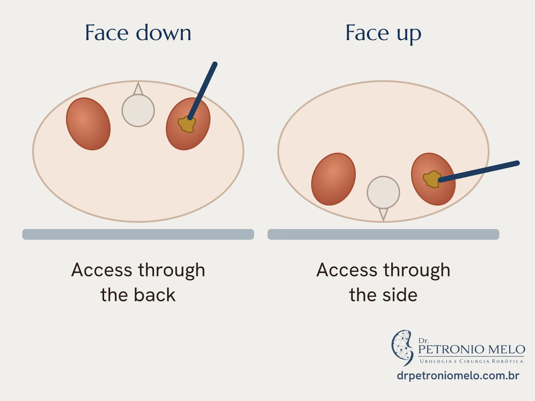
During my residency, we compared 393 operations in four positions: one face down and three face up. Success and serious complications were similar, and the complete face-up position had the shortest operating time.3 Today, I always operate face up, usually in the Barts flank-free position.
Larger and smaller instruments
Standard accesses are 24 to 30 Fr, and smaller ones were introduced for children and later for adults.1 In meta-analyses, mini-percutaneous surgery, at 12 to 22 Fr, removes the stone about as well as the standard approach, with less bleeding and fewer transfusions. The quality of this evidence is limited.1
For large, complex stones, a flexible nephroscope, or a ureteroscope through the urethra in the same operation, can help reach what is out of range of the rigid instrument.1
Without a nephrostomy tube
A nephrostomy is a tube that comes out through the skin, used after surgery in many cases. Operating without it, with the double J stent inside, is called tubeless surgery. It is associated with less pain and a shorter hospital stay, with low-certainty evidence.1 The European guideline recommends it in uncomplicated cases.1
Whenever possible, I do it this way. The final decision comes at the end of the operation, because it depends on bleeding, fragments, a planned second stage, blockage of the ureter and infection.1
Risks
A review of almost 12,000 patients gathered the complications of the operation.1
| Complication | Frequency |
|---|---|
| Fever | 10.8% |
| Transfusion | 7% |
| Chest complication | 1.5% |
| Sepsis | 0.5% |
| Injury to another organ | 0.4% |
| Embolization for bleeding | 0.4% |
| Death | 0.05% |
Untreated urinary infection, a tumor in the path of the access, suspected kidney cancer and pregnancy rule out the operation, and the plan for blood-thinning medicines is made in advance.1 Complex stones may need more than one access, with a higher risk of complications.1
My research in percutaneous surgery
During my residency, I was the first author of the study that gathered the service's experience: 1,066 percutaneous operations in 891 patients, from 2011 to 2016. Complications occurred in 14.9% of cases, and a CT scan the next day showed that the chance of being fragment-free fell from 87.9% in the simplest cases to 24.3% in the most complex.2 This figure helps us talk before surgery about the possibility of more than one procedure.
How I approach it
In my practice, I study the CT scan before deciding on the access. The patient is always face up, usually in the Barts flank-free position. Whenever possible, I operate without a nephrostomy tube, and the double J stent stays inside. The hospital stay is 24 hours. Before surgery, we talk about the chance of needing a second procedure, which depends on how complex the stone is.
When to see a urologist
- If you have fever or chills after surgery
- If your urine has a lot of blood or clots
- If severe pain does not improve with medicine
- If you are short of breath
- If a test shows a large kidney stone, even without pain
Frequently asked questions
When is percutaneous surgery recommended?+
It is the first option for kidney stones above 2 cm.1 It may also be used for smaller stones in the lower pole of the kidney and in selected cases of large, impacted stones in the upper ureter.1
Is the operation done lying face down?+
It can be done lying face down or face up, with equivalent results.1 I always operate with the patient face up, usually in the Barts flank-free position.
Will I leave with a drain in my back?+
Whenever possible, no. In my practice, I operate without a nephrostomy tube, leaving only the double J stent. The European guideline recommends this approach in uncomplicated cases.1 Bleeding, fragments or a planned second stage can change this.1
How long will I stay in hospital?+
In my practice, 24 hours.
What are the risks?+
In a review of almost 12,000 patients, there was fever in 10.8%, transfusion in 7%, a chest complication in 1.5%, sepsis in 0.5%, injury to another organ in 0.4% and death in 0.05%.1
Does one operation solve it?+
Often, but not always. In the study that gathered 1,066 operations from the service where I did my residency, the chance of being fragment-free fell from 87.9% in the simplest cases to 24.3% in the most complex.2
References
- EAU Guidelines on Urolithiasis. European Association of Urology; 2026. Guidelines chapter. EAU
- de Souza Melo PA, Vicentini FC, Beraldi AA, et al. Outcomes of more than 1 000 percutaneous nephrolithotomies and validation of Guy's stone score. BJU Int. 2018;121(4):640-646. PubMed
- Melo PAS, Vicentini FC, Perrella R, et al. Comparative study of percutaneous nephrolithotomy performed in the traditional prone position and in three different supine positions. Int Braz J Urol. 2019;45(1):108-117. PubMed
Would you like to talk about your case?
In the consultation, I go over your tests and your priorities.
Book a consultation