
Quick answer
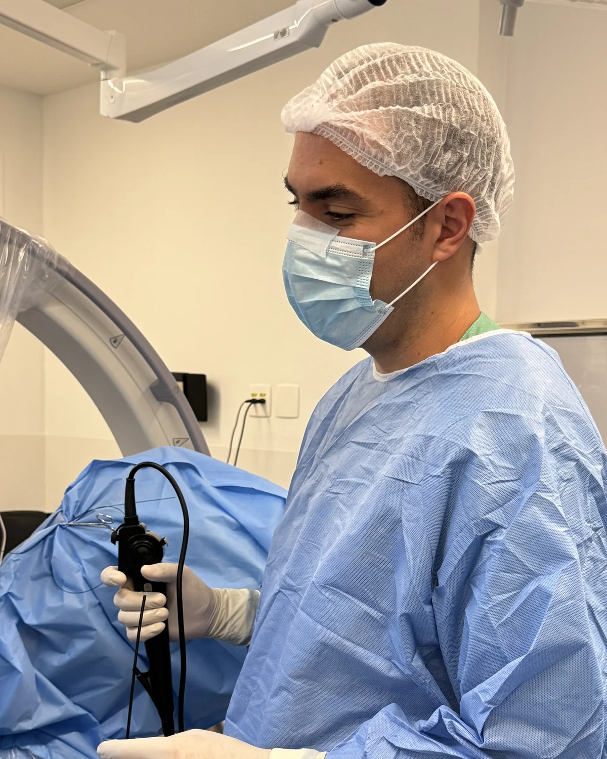
Flexible ureteroscopy is surgery without cuts. A thin scope goes in through the urethra, moves up the ureter and reaches the stone, which is broken up with a laser.1 It is used for stones in the ureter and in the kidney.1 In my practice, it is done under general anesthesia, the hospital stay is under 12 hours and I usually leave a double J stent for 5 days.
How it is done
- AnesthesiaIn my practice, the operation is done under general anesthesia.
- EntryThe scope goes in through the urethra and passes through the bladder, with no cut in the skin. A guidewire marks the way and adds safety.1
- Going upThe ureteroscope moves up the ureter to the stone. Its flexible tip bends and reaches the calyces of the kidney. A thin sheath often helps with going in and out and keeps the pressure inside the kidney low.1
- LaserA fiber passes through the scope, and the laser breaks the stone into fragments or into dust.1
- RemovalThe larger fragments come out with a nitinol basket or with forceps, always under direct vision.1
- EndIn my practice, I usually leave a double J stent, and the hospital stay is under 12 hours.
The laser and fluoroscopy
The European guideline recommends the holmium or thulium laser for flexible ureteroscopy.1 The laser turns energy into heat. That is why the operation needs continuous irrigation and control of the power and firing time.1
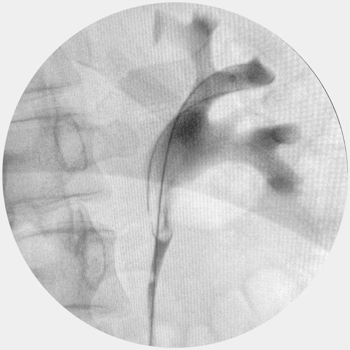
Fluoroscopy must be available in the room.1 With it, the team follows the path of the scope, and contrast outlines the inside of the kidney.
Who it is for
- Stones in the ureter. Ureteroscopy is more likely than shock waves to clear the stone in a single procedure.1
- Kidney stones, including in the lower pole.1
- Severe obesity, where it is the first option.1
- People who take blood-thinning medicines and cannot stop them, assessed case by case.1
- Stones above 2 cm, when percutaneous surgery is not an option, with a higher chance of needing another procedure.1
For kidney stones above 2 cm, one review showed 91% stone-free, with an average of 1.45 procedures per patient.1 The anatomy of the lower pole can make access harder.1
The double J stent after surgery
In my practice, I usually leave the double J stent: 5 days when surgery goes well, and longer if needed. It matters even more when there is injury to the ureter, fragments, bleeding, perforation or infection.1 If the scope cannot pass through the ureter on the first attempt, placing the stent and operating seven to fourteen days later is an alternative.1 For the discomfort and removal, see the double J stent page.
Risks
Complications occur in 4% to 25% of cases, and most are mild and do not require another procedure.1
| Complication | Frequency in the European guideline |
|---|---|
| Severe infection (sepsis) | Up to 5%1 |
| Narrowing of the ureter | 3% to 4.9%1 |
| Serious injury with rupture of the ureter | 0.04% to 0.9%1 |
A positive urine culture before surgery, earlier perforations, other illnesses and long operations raise the risk. The guideline advises keeping the operation under 90 minutes and treating infection beforehand.1
How I approach it
In my practice, I explain the plan before surgery and what may change during it, such as leaving the stent for longer or needing a second stage for large stones. Discharge comes in under 12 hours. Afterwards, we talk about the stone analysis and prevention.
When to see a urologist
- If you have fever or chills after surgery
- If severe pain does not improve with medicine
- If you cannot urinate
- If the blood in your urine becomes heavy or has clots
- If the agreed date for removing the stent is approaching
Frequently asked questions
Does laser surgery for kidney stones involve a cut?+
No. The scope goes in through the urethra, passes through the bladder and moves up the ureter to the stone.1
How long will I stay in hospital?+
In my practice, the stay is under 12 hours.
Is general anesthesia needed?+
In my practice, yes. The European guideline also says most operations are done under general anesthesia, although other types are possible.1
Will I have a double J stent after surgery?+
In my practice, usually yes. When surgery goes well, it stays for 5 days, and it may stay longer if needed.
Does it work for large stones?+
Above 2 cm, percutaneous surgery is the first option. Ureteroscopy is used when percutaneous surgery is not possible, with a higher chance of needing a second procedure.1
What are the risks?+
Complications occur in 4% to 25% of cases, and most are mild. Severe infection can occur in up to 5%, narrowing of the ureter in 3% to 4.9% and serious injury to the ureter in 0.04% to 0.9%.1
Does the stone go away completely?+
The goal is to remove the whole stone. With large stones, a fragment may remain or another procedure may be needed: for kidney stones above 2 cm, one review showed an average of 1.45 procedures per patient.1
References
- EAU Guidelines on Urolithiasis. European Association of Urology; 2026. Guidelines chapter. EAU
Would you like to talk about your case?
In the consultation, I go over your tests and your priorities.
Book a consultation