

Quick answer
PSMA PET is an imaging test that looks for prostate cancer cells in the body. The European guideline uses it in higher-risk cases, before treatment, and when PSA rises after surgery or radiotherapy.1,2 The higher the PSA, the greater the chance of finding the disease.5 A negative scan does not exclude the disease.1
What the scan is
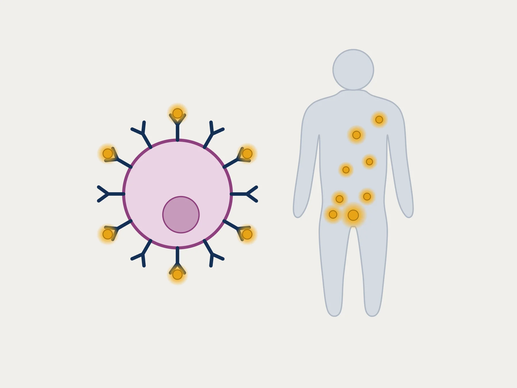
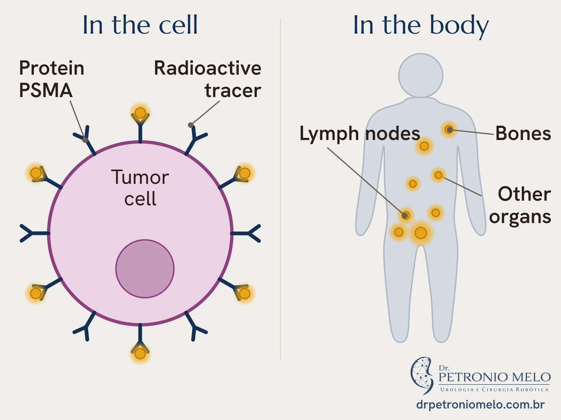
PET is an imaging test that shows where a radioactive substance has collected in the body. In PSMA PET, this substance binds to the PSMA protein and marks places where there may be tumor. The image is combined with a CT scan.1,7
MRI looks at the prostate in detail. PSMA PET looks for the disease in lymph nodes, bones and other organs.1
When the guideline recommends it
| Situation | What the European guideline advises |
|---|---|
| Low or favorable intermediate risk | No additional imaging for staging1 |
| Unfavorable intermediate risk | PSMA PET, when available, or CT with a bone scan1 |
| High risk or locally advanced disease | PSMA PET, when available, or CT with a bone scan1 |
| PSA that did not become undetectable after surgery | Offer the scan if PSA is rising and the result may change the decision2 |
| PSA that rose again after surgery | Do the scan if PSA goes above 0.2 ng/mL and the result may change the decision2 |
Before treatment
The proPSMA study randomly assigned 302 men with high-risk cancer to PSMA PET or conventional imaging. Accuracy was 92% with PSMA PET and 65% with CT and a bone scan.3
Its main use is staging, and the scan can also point to where to biopsy.1 In the PRIMARY study, with 291 men, adding PSMA PET to MRI raised sensitivity from 83% to 97%, while specificity fell from 53% to 40%.6
When PSA rises after treatment
PSMA PET helps show where the disease is, and the chance of finding it follows the PSA value:5
| PSA before the scan | Positive scan |
|---|---|
| Below 0.2 ng/mL | 33% |
| 0.2 to 0.49 ng/mL | 45% |
| 0.5 to 0.99 ng/mL | 59% |
| 1 to 1.99 ng/mL | 75% |
| 2 ng/mL or more | 95% |
In a study of 635 men, a positive image was confirmed as cancer in 84% of the cases with tissue analysis.4 In 323 men, the scan changed the treatment plan in 62%.2
How it is done
Limits of the scan
The scan does not see lesions smaller than about 5 mm.1 In a study of men who had surgery, it detected 40% of the cases with involved lymph nodes, with a specificity of 95%.1 That is why a negative result lowers the chance of distant disease without ruling it out.
PSMA also appears in other tumors and in benign changes, which can cause false positives.1 When a finding changes the plan, it is assessed together with the other tests.
How I approach it
In my practice, I order a PSMA PET when the result may change the decision. After surgery, I confirm that PSA is rising before ordering the scan. I read the report together with the PSA value and the surgical result, and only then do we talk about treatment.
When to see a urologist
- If your PSA rises after surgery or radiotherapy
- If you have been diagnosed and want to know whether you need the scan
- If your PSMA PET report shows a finding you did not understand
Frequently asked questions
What is a PSMA PET scan for?+
It looks for prostate cancer cells in the body. The European guideline uses it to assess the extent of the disease in higher-risk cases and to locate the tumor when PSA rises after treatment.1,2
At what PSA level does PSMA PET find the cancer?+
The higher the PSA, the greater the chance. In a review of 37 articles, the scan was positive in 33% of men with a PSA below 0.2 ng/mL and in 95% of men with a PSA of 2 ng/mL or more.5
Does a negative PSMA PET mean I do not have cancer?+
No. Very small lesions can be missed, because the scan sees down to about 5 mm.1 The result is read together with your PSA and the rest of your case.
Does PSMA PET replace MRI or biopsy?+
No. MRI is still the test that guides the biopsy, and the biopsy is the test that confirms cancer.1
Does every man with prostate cancer need the scan?+
No. In low-risk and favorable intermediate-risk disease, the European guideline advises against additional imaging for staging.1
Is the scan safe?+
The scan uses a radioactive substance injected into a vein. In a study of 635 men, there were no serious adverse events linked to it.4
References
- EAU-EANM-ESTRO-ESUR-ISUP-SIOG Guidelines on Prostate Cancer. European Association of Urology; 2026. Diagnostic evaluation chapter. EAU
- EAU-EANM-ESTRO-ESUR-ISUP-SIOG Guidelines on Prostate Cancer. European Association of Urology; 2026. Treatment chapter. EAU
- Hofman MS, Lawrentschuk N, Francis RJ, et al. Prostate-specific membrane antigen PET-CT in patients with high-risk prostate cancer before curative-intent surgery or radiotherapy (proPSMA): a prospective, randomised, multicentre study. Lancet. 2020;395(10231):1208-1216. PubMed
- Fendler WP, Calais J, Eiber M, et al. Assessment of 68Ga-PSMA-11 PET Accuracy in Localizing Recurrent Prostate Cancer: A Prospective Single-Arm Clinical Trial. JAMA Oncol. 2019;5(6):856-863. PubMed
- Perera M, Papa N, Roberts M, et al. Gallium-68 Prostate-specific Membrane Antigen Positron Emission Tomography in Advanced Prostate Cancer-Updated Diagnostic Utility, Sensitivity, Specificity, and Distribution of Prostate-specific Membrane Antigen-avid Lesions: A Systematic Review and Meta-analysis. Eur Urol. 2020;77(4):403-417. PubMed
- Emmett L, Buteau J, Papa N, et al. The Additive Diagnostic Value of Prostate-specific Membrane Antigen Positron Emission Tomography Computed Tomography to Multiparametric Magnetic Resonance Imaging Triage in the Diagnosis of Prostate Cancer (PRIMARY): A Prospective Multicentre Study. Eur Urol. 2021;80(6):682-689. PubMed
- Fendler WP, Eiber M, Beheshti M, et al. PSMA PET/CT: joint EANM procedure guideline/SNMMI procedure standard for prostate cancer imaging 2.0. Eur J Nucl Med Mol Imaging. 2023;50(5):1466-1486. PubMed
Would you like to talk about your case?
In the consultation, I go over your tests and your priorities.
Book a consultation